Doppler ultrasonography is an important diagnostic technique that evaluates blood flow inside the body of the fetus and mother using sound waves. This non-invasive imaging technique utilizes sound waves to examine blood flow, helping doctors monitor the developing fetus and detect potential complications. It can be used to assess blood flow in various parts of the body, including arteries, veins, and the heart.
Doppler Ultrasound: What Is It Used For?
- Monitoring Blood Flow: The placenta, umbilical cord, and fetal organs’ blood flow are all measured with the help of Doppler ultrasound. This aids fetal medicine specialists in detecting anomalies or disturbances in blood flow that can impact the growth and well-being of a developing fetus.
- Assessing Fetal Well-being: Doppler ultrasound helps in evaluating the overall well-being of the fetus by examining its cardiovascular system. Variations in blood flow patterns can be a sign of possible issues such as fetal discomfort, which can be managed and intervened upon promptly.
- Identifying High-Risk Pregnancies: Doppler ultrasound is instrumental in identifying pregnancies at high risk for complications, such as preeclampsia or intrauterine growth restriction (IUGR). When these disorders are identified early on, medical professionals can take the necessary precautions to protect the mother’s and the unborn child’s health.
What to Expect During a Doppler Ultrasound?
- Non-Invasiveness:
Doppler ultrasound is a non-invasive procedure, similar to the routine pregnancy ultrasound study. It does not involve any needles or surgical instruments. It is a safe and painless technique that poses no risk to the mother and the baby.
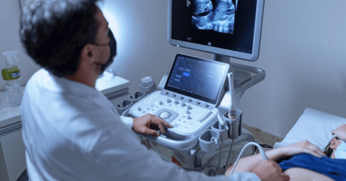
- Procedure Overview:
During the examination, a gel is applied to the mother’s abdomen to facilitate the transmission of sound waves. A transducer, a handheld device, is then moved over the abdomen to capture images of blood flow in the targeted areas.
- Duration of the Procedure:
Doppler ultrasounds usually take 15-20 minutes to complete. This is a fairly short procedure. Depending on the complexity of the examination and what particular areas are being assessed, the duration may change.
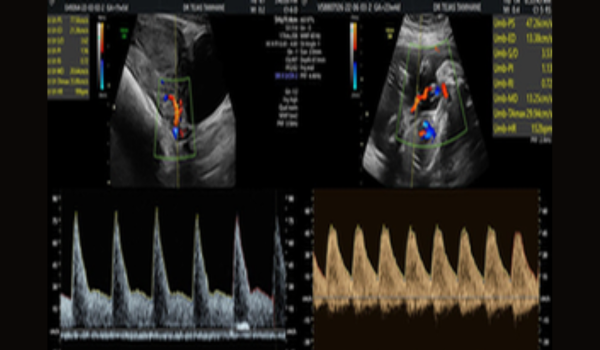
Results and Interpretation
- Normal Blood Flow Patterns:
Normal Doppler ultrasonography readings indicate a healthy blood flow pattern in the umbilical cord and some important fetal organs. This implies that sufficient oxygen and nutrition are being given to the fetus, enabling healthy growth and development.
- Abnormal Findings:
Issues like restricted blood flow that may be a sign of preeclampsia or IUGR can be found on Doppler ultrasound readings. When these irregularities are promptly identified, medical professionals can take the necessary steps to address them, such as closely monitoring or arranging an early birth.
- Decision-Making:
Equipped with the results of a Doppler ultrasound, medical professionals and pregnant parents can make well-informed and cooperative decisions. This may involve adjustments to the pregnancy management plan, more frequent monitoring, or specialized care to address any identified concerns.
In Conclusion
Fetal medicine experts use Feto-maternal Doppler ultrasound to identify heart and blood vessel (cardiovascular) complications. The procedure shows the direction and speed of blood moving through the arteries and veins. Appropriate interpretation of the Doppler ultrasound study is very important to avoid any complications due to abnormal blood flow.
Doppler Ultrasound in Pune
Dr Tejas Tamhane, one of the most renowned fetal medicine specialists in Pune, has years of experience and expertise in doing Doppler scans. So, if you are looking for a Doppler Test in Pune, contact his team at Precious Clinic.